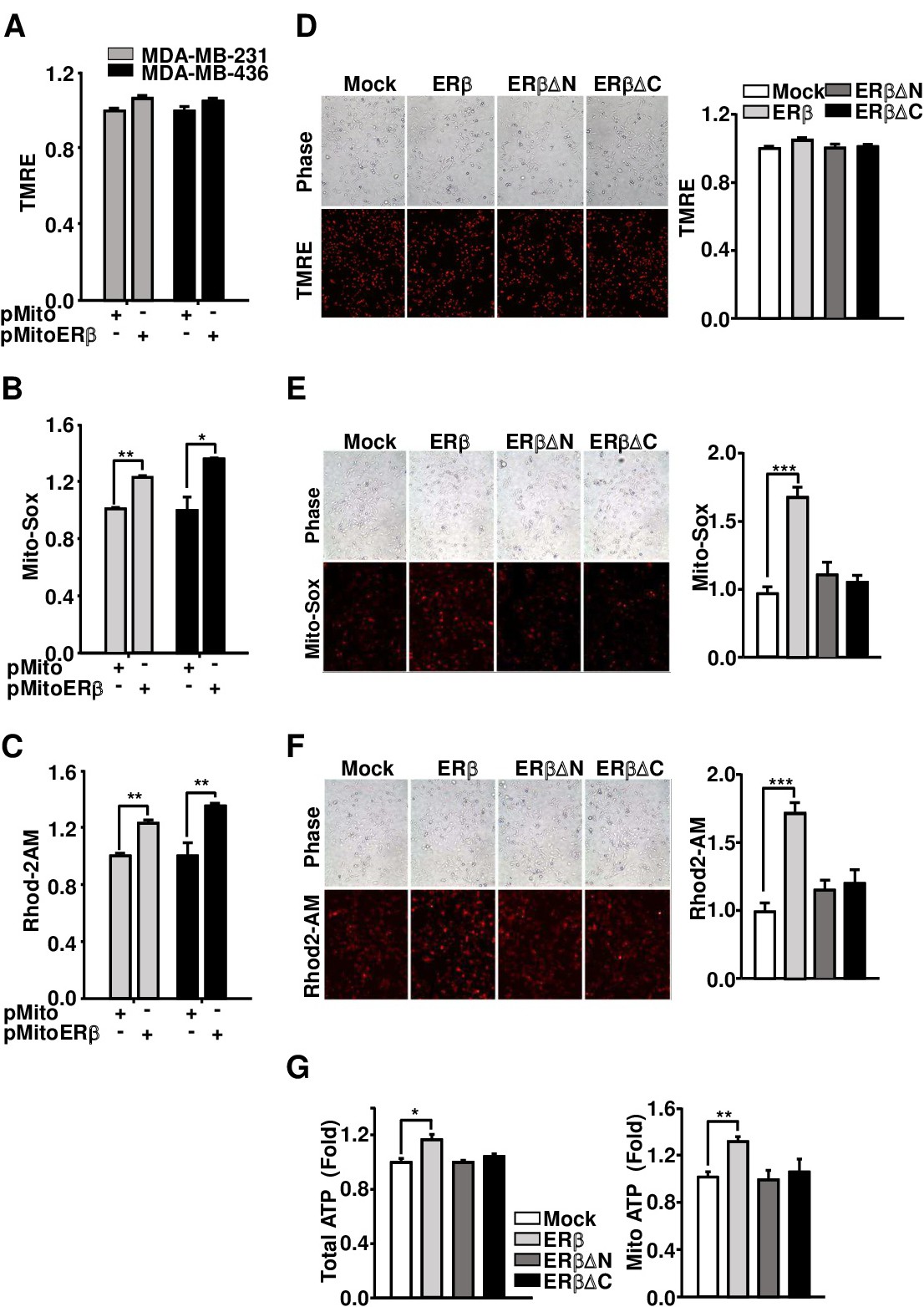
Fig. 6. mitoERβ improves the mitochondrial function in TNBC MDA-MB-231 and -436 cells. A-C. Mitochondrial membrane potential (A), ROS levels (B), and calcium levels (C) were measured in mock- and pHA-mito-ERβ-transfected TNBC MDA-MB-231 and MDA-MB-436 cells. D-F. MDA-MB-231 cells were transfected with mock, ERβ, and ERβ deleted mutants as indicated, and the cells were stained with TMRE (D), Mito-Sox (E), and Rhod-2AM (F). Representative fluorescent microscope images are shown, with quantified data shown as a graph in the right panel. G. MDA-MB-231 cells were transfected with mock, pHA-ERβ, pHA-ERβ△N, and pHA-ERβ△C, and total and mitochondrial ATP levels were measured by quantifying the luciferase-catalyzed ATP-dependent oxidation of luciferin.